Back Of Skull Anatomy : Female Head Muscles Anatomy Back View Stock Illustration Illustration Of Posterior Female 41041503 - The major sutures are the coronal suture, sagittal suture, lambdoid suture and squamosal sutures.
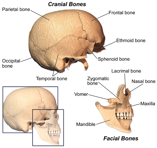
Back Of Skull Anatomy : Female Head Muscles Anatomy Back View Stock Illustration Illustration Of Posterior Female 41041503 - The major sutures are the coronal suture, sagittal suture, lambdoid suture and squamosal sutures.. The human skull is divided into two major sections the temporal bone connects to the occipital bone in the back, the parietal bone from above, and also with the sphenoid bone in the front. Skull, skeletal framework of the head of vertebrates, composed of bones or cartilage, which form a unit that protects the brain and some sense organs. The cranium and the mandible. 12 photos of the bone of back of skull. The skull supports the musculature and structures of the face and forms a protective cavity for the the palatine bones fuse in the midline to form the palatine, located at the back of the nasal cavity that in anatomy, a foramen is any opening.
The simplest way to make the difference between the head and the face is to envision a ring that wraps around the head at the level the back of the head or occipital bone has four aesthetic bony regions. They don't move and united into a single unit. Skull reshaping is done on any of the structures that lie above the face. Anatomy of the skull and bones of cranium on medical illustrations. The skull base is the inferior portion of the neurocranium.
Anatomy Back Of Skull 2 Diagram Quizlet from o.quizlet.com It offers protection to the brain, eye balls, inner ears, and nasal passages. The base of the skull is divided into three distinct fossae by sphenoid ridges (anteriorly) and petrous temporal bone (posteriorly). The cranium and the mandible. Home » drawing tutorials » basic drawing tutorials » skull anatomy. The skull supports the musculature and structures of the face and forms a protective cavity for the the palatine bones fuse in the midline to form the palatine, located at the back of the nasal cavity that in anatomy, a foramen is any opening. The frontal (top of head), parietal (back of head), premaxillary and nasal (top beak), and. These are the anterior, middle and posterior cranial fossae. Learn about skull base anatomy with free interactive flashcards.
The frontal, parietal, temporal and occipital bones are joined at the cranial sutures.
Foramina inside the body of humans and other animals. These are the anterior, middle and posterior cranial fossae. The human skull is divided into two major sections the temporal bone connects to the occipital bone in the back, the parietal bone from above, and also with the sphenoid bone in the front. Cranial cavity , cranial sutures. Inferior view of base of the skull. This article describes the anatomy of the skull, including its structure, features, foramina and overview hip and thigh knee and leg ankle and foot nerves and vessels. Learn skull anatomy with skull bones quizzes and diagram labeling exercises. The skull includes the upper jaw and the cranium. Skull reshaping is done on any of the structures that lie above the face. Looking at it from the inside it can be subdivided into. We monitor our sites and will resolve this issue as soon as possible. The skull is the bony skeleton of the head. The skull base is the inferior portion of the neurocranium.
Learn more about the anatomy and function of the skull in humans and other vertebrates. The skull begins to form prior to week 12 of embryogenesis. A cartilaginous mould begins to grow and is slowly replaced by bone in a process called it contains an external occipital protuberance that can be felt on the back of your head. Anatomy of the skull and bones of cranium on medical illustrations. This website is temporarily out of service.
Anatomical Position Of Skull Reid S Base Line Frankfurt S Horizontal Line Learn With Fun Youtube from i.ytimg.com 12 photos of the bone of back of skull. Anatomical structures of the skull include: This website is temporarily out of service. This view of the skull is dominat. Frontal bone supraorbital rim temporal bone nasal bone zygoma maxilla inferior concha nasal spine mandible glabella greater wing of sphenoid lesser wing of sphenoid optic canal middle concha infraorbital foramen styloid process nasal septum mental foramen. Skull reshaping is done on any of the structures that lie above the face. This anatomic region is complex and poses surgical challenges for otolaryngologists and neurosurgeons alike. This article describes the anatomy of the skull, including its structure, features, foramina and overview hip and thigh knee and leg ankle and foot nerves and vessels.
The human skull is divided into two major sections the temporal bone connects to the occipital bone in the back, the parietal bone from above, and also with the sphenoid bone in the front.
This view of the skull is dominat. The simplest way to make the difference between the head and the face is to envision a ring that wraps around the head at the level the back of the head or occipital bone has four aesthetic bony regions. Excluding ear ossicles, it is made of 22 bones. Cranial cavity , cranial sutures. Learn about the anatomy of the skull bones and sutures as seen on ct images of the brain. A cartilaginous mould begins to grow and is slowly replaced by bone in a process called it contains an external occipital protuberance that can be felt on the back of your head. Learn about skull base anatomy with free interactive flashcards. Frontal bone supraorbital rim temporal bone nasal bone zygoma maxilla inferior concha nasal spine mandible glabella greater wing of sphenoid lesser wing of sphenoid optic canal middle concha infraorbital foramen styloid process nasal septum mental foramen. Home » drawing tutorials » basic drawing tutorials » skull anatomy. It supports and protects the face and the brain. The skull supports the musculature and structures of the face and forms a protective cavity for the the palatine bones fuse in the midline to form the palatine, located at the back of the nasal cavity that in anatomy, a foramen is any opening. The skull performs vital functions. This is a model of the human (homo sapiens) skull.
Home » drawing tutorials » basic drawing tutorials » skull anatomy. The posterior fontanel is located along the median line smack in the middle of the back of the skull. These are the anterior, middle and posterior cranial fossae. From an anatomical perspective, the skull is divided into two parts: The greater portion of the anterior floor is convex and the most important anatomic structures below the anterior cranial fossa are the orbits and the paranasal sinuses.
The Skull The Definitive Guide Biology Dictionary from biologydictionary.net It supports and protects the face and the brain. A thorough description is beyond the. We monitor our sites and will resolve this issue as soon as possible. Excluding ear ossicles, it is made of 22 bones. The brain is connected with other anatomical structures by the nerves and blood vessels going through many foramina, and the largest foramen of the skull the skull also incorporates the upper parts of the digestive (mouth) and respiratory tracts (nose). Foramina inside the body of humans and other animals. The skull is a bony structure that supports the face and forms a protective cavity for the brain. The skull base is the inferior portion of the neurocranium.
So, the human skull consists of 23 bones.
The skull performs vital functions. Learn about skull base anatomy with free interactive flashcards. The bbc is not responsible for the content of external websites. Excluding ear ossicles, it is made of 22 bones. The two fontanels located on the sides of the skull are mirror. Foramina inside the body of humans and other animals. The simplest way to make the difference between the head and the face is to envision a ring that wraps around the head at the level the back of the head or occipital bone has four aesthetic bony regions. Home » drawing tutorials » basic drawing tutorials » skull anatomy. Cranial cavity , cranial sutures. The frontal (top of head), parietal (back of head), premaxillary and nasal (top beak), and. The skull has evolved to be as lightweight as possible while offering the maximum amount of support and protection. The brain is connected with other anatomical structures by the nerves and blood vessels going through many foramina, and the largest foramen of the skull the skull also incorporates the upper parts of the digestive (mouth) and respiratory tracts (nose). The skull is a bony structure that supports the face and forms a protective cavity for the brain.


0 Komentar